
3D Cone Beam Imaging in Exton, PA: Clear 3D Dental Views
Cone beam imaging in Exton, Pennsylvania, supports precise diagnosis and treatment planning across many areas of dentistry. This advanced 3D dental X-ray offers a detailed look at teeth, bone, nerves, and sinus structures in a single scan, helping patients and clinicians make informed decisions.
Cone Beam Imaging Explained
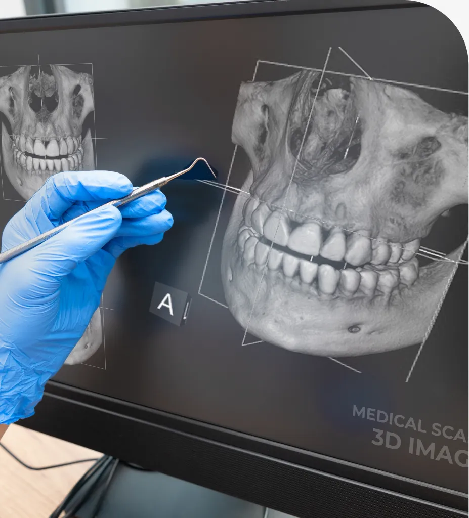
Cone beam imaging, also called cone beam computed tomography (CBCT), is a specialized form of dental CT that creates a three-dimensional image of your mouth and jaws. Unlike traditional two-dimensional X-rays, a CBCT dental scan rotates around your head and captures hundreds of images in seconds. Software then reconstructs those images into a 3D model that can be viewed from any angle.
Dr. Santosh Mittal uses CBCT to assess dental implants, impacted teeth, root canal anatomy, jaw joints, airway and sinus concerns, bone quality, and facial trauma. The scan reveals details that standard X-rays cannot show, which can reduce surprises during treatment and improve accuracy.
Benefits of Cone Beam Imaging
- Comprehensive 3D detail for precise diagnosis and planning.
- Targeted imaging of the area of interest to focus on relevant anatomy.
- Efficient scan time, often under 20 seconds for image capture.
- Improved implant planning with accurate bone measurements and nerve mapping.
- Better visualization of root canals, fractures, and complex tooth anatomy.
- Evaluation of jaw joints, sinuses, and airway in one scan.
- Fewer repeat images when the 3D view answers key questions up front.
How Cone Beam Imaging Works
During a CBCT appointment, the area to scan is selected based on your clinical needs. You may sit or stand in the unit while resting your chin or forehead against supports. The scanner rotates in a circle without touching you, and image capture usually takes 10 to 20 seconds. The system then reconstructs a high-resolution 3D model for review.
Because precise positioning matters, you will be asked to remain still and to remove glasses, earrings, removable dental appliances, and any metal objects that could interfere with the image.
What to Expect
Most patients describe cone beam imaging as quick and comfortable. There are no injections, impressions, or claustrophobic tunnels. The radiation dose is typically lower than a medical CT scan and higher than a few standard dental X-rays. As with all imaging, the ALARA principle (“as low as reasonably achievable”) guides decision-making to ensure the scan is justified and optimized for your needs.
Pregnant patients should tell our team before any X-rays or scans. Alternative timing or protective measures may be considered. Results are available immediately, and our dentist can review the images with you to explain findings, outline options, and plan the next steps.
The Cone Beam Imaging Process & Benefits
The Cone Beam Imaging Process
- Initial review: Our dentist identifies why a 3D dental X-ray is helpful and selects the smallest necessary scan area.
- Positioning: You are seated or standing, guided into the correct position, and asked to remain still.
- Scan: Image capture takes seconds; there is no discomfort.
- Interpretation: Images are reconstructed into 3D views for clinical analysis and treatment planning.
How Cone Beam Imaging Can Help You
- Implants: Accurate planning reduces surgical risks and aligns implant placement with restorative goals.
- Root canals: High-definition views reveal extra canals, fractures, or hidden infections.
- Orthodontics: Jaw relationships, impacted teeth, and airway can be assessed in detail.
- Oral surgery: Wisdom teeth proximity to nerves and sinus anatomy are evaluated before procedures.
Frequently Asked Questions About Cone Beam Imaging
Ready for 3D Cone Beam Imaging : Clear 3D Dental Views?
Our team provides expert 3d cone beam imaging : clear 3d dental views with compassionate, personalized care.